
Spatiotemporal Omics: The Next Frontier in Molecular Biology
Why Spatial and Temporal Contexts Are Inseparable?
Among the most dynamic and rapidly expanding domains in omics research, spatial omics enables the investigation of DNA, RNA, proteins, metabolites, and other small molecules within the context of tissue architecture. This preservation of spatial molecular information facilitates a more holistic understanding of cellular behavior and intercellular communication. The discipline initially focused on two-dimensional (2D) spatial omics across the x and y axes on one plane. Recently, the field has advanced toward the third dimension (3D), which expands the analysis into z-axis or 3D space. Although still emerging, this allows for a holistic understanding of molecular expression within tissues.
While both 2D and 3D spatial omics preserve location-specific information, they provide only a single time-point view. To uncover the full complexity of biological processes, it is essential to monitor dynamic changes in biomolecules within the same living tissue sample over time. This is the very goal that spatiotemporal omics technologies are developed to attain.
Top Technologies Driving Spatiotemporal Monitoring
The past few years have seen significant advances in single-cell spatial and temporal omics technologies. These developments include the application and refinement of previously established live imaging and spatial omics techniques for monitoring biological processes in 2D and 3D. The combination of these methods allows researchers to capture dynamic molecular changes with high resolution, enabling a more integrated understanding of cellular behavior across both time and space. In this section, we offer a concise overview of the technologies that support detailed spatiotemporal analysis of single-cell omics data.
(i) In situ hybridization (ISH):
ISH is a targeted spatial transcriptomics technique that enables the detection and localization of specific RNA molecules within intact tissue sections. Offering subcellular resolution, ISH has proven especially valuable for uncovering spatial and temporal gene expression dynamics in complex biological systems such as organoids and developing tissues.
For example, ISH has been effectively used to study engineered gene expression patterns in organoids, where transcript activation and suppression were modulated to mimic developmental signaling pathways. Through activation of the Sonic hedgehog pathway and the integration of ISH with complementary spatial and single-cell transcriptomic approaches, researchers were able to localize regions with distinct gene expression and gain insights into the regulation of genes at different stages of activation.
In another study, ISH was combined with cellular barcoding to allow the simultaneous analysis of gene expression, single-cell lineage, and spatial organization within the same tissue. This approach enabled the identification of clonal structures and gene expression patterns in mouse embryonic stem cells and Drosophila brains across the period of neural development.
(ii) In situ sequencing (ISS):
ISS is a targeted spatial transcriptomics technique that sequences RNA molecules directly within tissue sections, allowing for the simultaneous detection of a large number of genes and offering a higher multiplexing capacity compared to traditional ISH. This method has proven particularly valuable for investigating complex biological phenomena, such as A-to-I RNA editing.
A notable advancement in this area is the development of TEMPOmap, a method designed to uncover subcellular RNA profiles across time and space at the single-cell level. It was employed to integrate metabolic labeling of RNA transcripts, rolling circle amplification (RCA), and 3D imaging within a hydrogel–cell scaffold. High-resolution spatial mapping was accomplished using computational tools such as 3D fast Fourier transforms and MATLAB-based analyses. Additionally, researchers monitored transcription, nuclear export, and decay rates across thousands of genes through pulse-chase labeling. This approach revealed that mRNA regulation varied across different stages of the RNA life cycle and distinct phases of the cell cycle.
ISS has also been integrated with high-throughput clonal tracking to assess cell phenotypes and trace clonal relationships in complex tissues like the developing mouse brain. In this context, individual progenitor cells were barcoded and analyzed using ISS alongside single-cell RNA sequencing. This combined approach enabled in situ clonal tracing and gene expression analysis, leading to the identification of fate-restricted progenitor cells in the mouse hippocampal neuroepithelium and revealing that microglia arise from a small population of primitive myeloid precursors.
(iii) Next-Generation Sequencing (NGS)-based spatial barcoding
Several studies have employed NGS-based spatial barcoding as a powerful tool for monitoring and tracking developmental changes at high resolution:
- Chen et al. used Stereo-seq to investigate spatial variations among cells and cell fate decisions in the developing dorsal midbrain. They constructed a mouse organogenesis spatiotemporal transcriptomic atlas (MOSTA), which captured transcriptional variations, their directionality, and kinetics throughout organogenesis. The dataset included 53 fixed sagittal sections from C57BL/6 mouse embryos spanning embryonic day (E) 9.5 to E16.5 in daily intervals.
- From a 3D perspective, Slide-seq was used by Sampath Kumar et al. to analyze entire mouse embryos at the onset of organogenesis. This technique enabled the generation of spatially resolved gene expression profiles across multiple developmental time points.
- In another study, Wang et al. utilized NGS-based spatial barcoding to construct 3D transcriptomic maps of developing Drosophila embryos, shedding light on the kinetics of gene expression in both 2D and 3D contexts during development.
(iv) Multiplex immunostaining
Multiplex immunostaining facilitates spatiotemporal monitoring by using multiple antibodies to simultaneously detect various proteins in living cells or tissues, often enhanced by bioorthogonal click chemistry techniques.
The method allows for multiplexed evaluation of changes in protein expression over time. This technique has been applied to both cells and organoids, leading to key discoveries. Some studies have also used immunostaining to study protein dynamics at the single-cell level, offering deeper insight into critical biological processes such as the cell cycle. These investigations have revealed new findings regarding intercellular proteomic heterogeneity and the role of cycling proteins in driving cell proliferation and contributing to oncogenesis.
Despite its strengths, multiplex immunostaining has limitations. Notably, it cannot profile the transcriptome or genome, and it relies heavily on the availability of antibodies.
(v) Bioelectronics
Bioelectronics offers an innovative approach to spatiotemporal monitoring by integrating electronic devices, such as open-mesh woven structures, into living tissues to record cellular activity over extended periods. These devices, like those used to track single-unit action potentials in mouse brains, seamlessly integrate with neural networks, minimizing immune responses and enabling long-term electrophysiological monitoring.
The integration of bioelectronics with techniques such as in situ RNA sequencing allows researchers to map both electrical activity and gene expression from the same cells, offering a multidimensional view of cellular function.
Methodological Approaches to Experimental Design
Depending on the complexity of the research question, three main experimental design frameworks are commonly used: single-sample, multi-sample, and continuous-sample designs. Each differs in how it structures sample collection and analysis to support targeted insights.
The continuous sample design is tailored for studies requiring molecular characterization along a continuous covariate, such as time or space. This framework involves collecting multiple samples from a subject at different time points or spatial locations, enabling the tracking of molecular changes over a continuum. For instance, profiling a tumor biopsy at various spatial coordinates or monitoring gene expression in a developing embryo over time falls under this design. It is particularly powerful for modeling temporal or spatial dependencies, allowing researchers to identify features that vary smoothly across time or space, such as spatially variable genes or temporally dynamic proteins. This design supports advanced analyses, like interpolation of missing data or realignment of coordinates across subjects, making it essential for studying dynamic processes in development, disease progression, or tissue architecture.
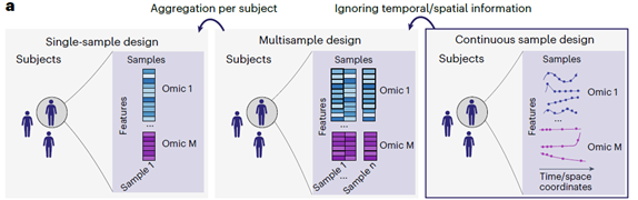
Adapted from: Velten B, et al. Nat Methods. 2023;20(10):1462–1474.
Strategies to Model Spatial and Temporal Dependencies
As continuous sampling becomes more common in dataset designs, a diverse set of analytical methods has been developed to account for and utilize the underlying spatial and temporal structures in the data. At the heart of these methods are modeling components that define the dependencies of a sample characteristic z at the time point t or spatial position x of the sample. Typically, z represents one/multiple observed omics features, such as an expression vector of genes or a compositional vector of microbial or cell type abundances.
Instead of modeling the temporal or spatial relationship of observed omic features, it can also be helpful to consider dependencies between time or space and an unobserved parameter that is derived from the observed omic features. Latent variable models are a well-known example of this strategy. They describe the link between observed measurements (e.g. feature abundances) to hidden variables (e.g. cell types, clusters, or low-dimensional embeddings). These models are often used for tasks like cell type deconvolution or dimension reduction in temporal or spatial applications.
Whether z represents an observed or latent sample characteristic, similar modeling strategies can be applied to describe dependencies between z and time or space. Alternative modeling methods can be grouped into two broad categories: regression-based and graph-based models.

Examples of regression-based modeling based on
Parametric regression (illustrated for a sigmoid function with parameters a and b)
Splines (composed of polynomial functions pn for disjoint consecutive intervals)
Gaussian Process (parametrized by a mean function μ and covariance function κ)
Adapted from: Velten B, et al. Nat Methods. 2023;20(10):1462–1474.
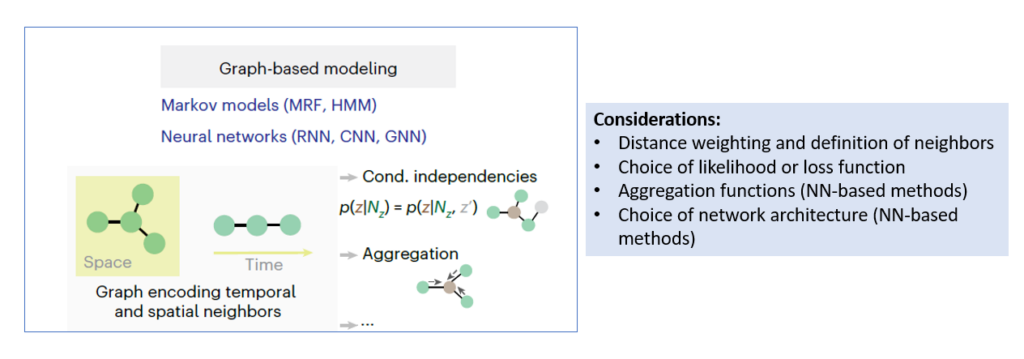
Examples of graph-based representations for space and time
Nodes denote samples with labels corresponding to measurements or model parameters
Edges encode temporal or spatial proximity
Adapted from: Velten B, et al. Nat Methods. 2023;20(10):1462–1474.
CNN: Convolutional neural network; cond.: conditional; GNN: Graph neural network; HMM: Hidden Markov model; MRF: Markov random field; NN: Neural network; RNN: Recurrent neural network
CNN: Convolutional neural network; cond.: conditional; GNN: Graph neural network; HMM: Hidden Markov model; MRF: Markov random field; NN: Neural network; RNN: Recurrent neural network
Selecting a suitable model class for a given application often involves balancing tradeoffs between computational scalability, model interpretability, and performance for a specific task. Some general recommendations include:
- Using regression-based models for small and low-dimensional datasets
- Using graph-based models when spatial or temporal dependencies are complex
- Using multivariate models that can share information across features and omic layers for large, high-dimensional datasets
Several approaches have been developed to jointly model temporal or spatial dependencies in multi-omic data. These methods often involve technology-specific normalization during preprocessing or incorporate modality-specific noise models, possibly aligning temporal or spatial dimensions across omic layers.
What Are We Gaining with Spatiotemporal Omics?
Spatiotemporal omics hold the potential to fundamentally reshape our understanding of key biological processes and provide answers to long-standing clinical questions. In this section, we briefly highlight several areas of medicine where this emerging approach could be particularly impactful.
(i) Detailed understanding about the pathomechanism of diseases:
One of the most impactful applications of spatiotemporal omics is its ability to uncover the intricate molecular foundations of diseases
- In cancer research, for example, spatial heterogeneity and temporal evolution pose significant challenges. Tumors are not uniform—distinct regions within the same tumor can behave differently and respond variably to therapy. Over time, these regions may shift, giving rise to treatment resistance, recurrence, or metastasis.
- In neurodegenerative diseases like Alzheimer’s protein aggregates such as amyloid plaques do not appear uniformly; their distribution varies across brain regions and progresses gradually. Mapping these patterns helps clarify how the disease spreads and disrupts brain function.
- Similarly, aging is not a uniform process. Different tissues, or even regions within the same organ, can age at different rates, resulting in spatially distinct molecular signatures.
Spatiotemporal omics provides the resolution and context needed to dissect these complex biological landscapes, offering new insights into how localized molecular changes drive disease onset, progression, and variability.
(ii) Enabling early detection and better risk profiling
Another important application of this approach lies in predicting the risk of metastasis and recurrence, which are critical factors in managing conditions like cancer. Spatiotemporal omics allows researchers to correlate the spatial context of these processes with clinical outcomes, helping identify regions within a tumor that may be more likely to spread or resist therapy. These insights can significantly refine existing prognostic models, providing clearer predictions around patient survival, treatment response, and disease progression.
In addition, this novel omics approach could help bridge histopathology and molecular biology by overlaying high-resolution molecular profiles onto tissue architecture. Such integration may significantly enhance current strategies for the early detection of diseases.
As these technologies advance toward clinical adoption, they hold the potential to transform routine diagnostics and prognostics, enabling more targeted, timely, and effective disease detection.
(iii) Bridging the gap to truly individualized care
The integration of spatiotemporal omics with advanced model systems like patient-derived tissues and organoids would create environments that better reflect human physiology compared to traditional animal models, which tend to be expensive, time-consuming, and less predictive of human responses. Through such profiling, clinicians and researchers can monitor how diseases evolve and respond to therapies over time, gaining valuable insight into drug metabolism, distribution, and adverse effects. This temporal resolution supports better treatment planning and helps bridge the gap between preclinical studies and real-world clinical outcomes.
Beyond therapeutic planning, these approaches could provide deeper insights into complex, heterogeneous conditions such as glioblastoma and lung cancer, where tumor behavior differs across regions. With spatially and temporally resolved molecular data, the technology would provide a solid foundation for personalized therapy planning and empower clinicians to make more informed, adaptive decisions tailored to each patient’s unique clinical profile.
Spatiotemporal omics marks a paradigm shift in how we study life at the molecular level. As the field evolves, there is an increasing emphasis on integrating complementary datasets and refining sampling strategies to capture critical transitions in space and time. Future success will depend on our ability to balance resolution, coverage, and relevance. The focus must be on collecting data not only comprehensively but also with precision and intent. This progress brings us closer to a future where biological systems can be observed and fully understood in full four-dimensional (4D) perspective.
References:
1. Velten B, Stegle O. Principles and challenges of modeling temporal and spatial omics data. Nat Methods. 2023;20(10):1462–1474. doi:10.1038/s41592-023-01992-y
2. Reynolds DE, Roh YH, Oh D, et al. Temporal and spatial omics technologies for 4D profiling. Nat Methods. Published online April 22, 2025. doi:10.1038/s41592-025-02683-6
3. Chu LX, Wang WJ, Gu XP, et al. Spatiotemporal multi-omics: exploring molecular landscapes in aging and regenerative medicine. Mil Med Res. 2024;11(1):31. doi:10.1186/s40779-024-00537-4
4. Zhang J, Yin J, Heng Y, et al. Spatiotemporal Omics-Refining the landscape of precision medicine. Life Med. 2022;1(2):84–102. doi:10.1093/lifemedi/lnac053
5. Zuo C, Zhu J, Zou J, et al. Unravelling tumour spatiotemporal heterogeneity using spatial multimodal data. Clin Transl Med. 2025;15(5):e70331. doi:10.1002/ctm2.70331
6. Chen A, Liao S, Cheng M, et al. Spatiotemporal transcriptomic atlas of mouse organogenesis. Cell. 2022;185(10):1777–1792. doi: 10.1016/j.cell.2022.04.003