
What’s Happening in Single-Cell-based Medical Research?
Six Studies Worth Your Time
In just a few years, single-cell sequencing has evolved from a niche innovation into a driving force in biomedical research. It allows scientists to examine the activity of individual cells in remarkable detail, revealing how they behave, communicate, and respond to disease. This high-resolution perspective is transforming how we study a wide range of conditions, from viral infections to cancer progression.
With each step forward, the field is having a more profound impact on the field of medicine. In this blog post, we highlight six standout studies published in recent years that exemplify this transformation. From identifying immune signatures in diverse populations to charting tumor heterogeneity with precision, each study showcases the remarkable potential of single-cell research to shape the future of diagnostics and therapies.
Whether you are a researcher, clinician, or curious reader, read on to discover how science is becoming more insightful by listening to every single cell.
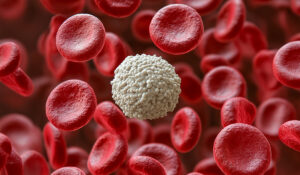
1. Peripheral blood immune responses in COVID-19
What happens in our blood when COVID-19 strikes? A 2021 study by Stephenson et al. in Nature Medicine dives into this question with a massive single-cell multi-omics analysis, profiling over 780,000 peripheral blood mononuclear cells (PBMCs) from 130 patients across a spectrum of COVID-19 severities, from asymptomatic to critical. Using single-cell RNA sequencing, surface proteome analysis, and T and B cell receptor profiling, the researchers uncovered how our immune system orchestrates its response to SARS-CoV-2, revealing potential therapeutic targets.
The study found that severe COVID-19 is marked by an expansion of nonclassical monocytes that engulf platelets and may replenish alveolar macrophages in the lungs, contributing to inflammation. Early hematopoietic stem cells showed a bias toward megakaryopoiesis, leading to increased platelet activation, a clue to the clotting issues seen in severe cases.

On the T cell front, severe disease was linked to clonally expanded CD8+ effector T cells and a higher ratio of effector to memory T cells, while mild cases featured more follicular helper T cells, suggesting a protective role. Interestingly, despite an increase in plasmablasts and plasma cells, symptomatic patients showed a marked decrease in IgA2 antibodies. These findings highlight the complex interplay of blood immune cells in COVID-19 and point to specific cellular components for future therapies.
2. A look at healthy and fibrotic human liver at single-cell resolution
This groundbreaking study by Watson et al. in Nature Communications used multiplexed error-robust fluorescent in situ hybridization (MERFISH) and single-nucleus RNA sequencing (snRNA-seq) to map the human liver’s cellular landscape at single-cell resolution. Focusing on both healthy and fibrotic livers, the researchers threw light on the spatial organization of hepatocytes, macrophages, and hepatic stellate cells (HSCs) within hepatic lobules and distinct hepatocyte sub-populations that expand with fibrotic injury.
The integration of MERFISH spatial data with snRNA-seq transcriptomes enabled the identification of receptor-ligand interactions enriched in distinct zones of liver. Specific pairs were localized to different zones, revealing spatially defined signaling between hepatocytes and non-parenchymal cells. These interactions form the basis of the liver’s spatial division of labor, supporting its metabolic and immune functions.
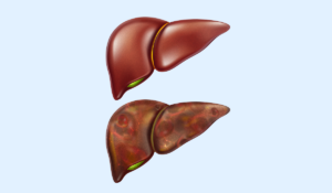
Furthermore, the altered gene expression in fibrotic hepatocytes reflects the transition to a pathological state, potentially contributing to liver dysfunction and disease progression. The disruption of zonal gene expression patterns in fibrosis suggests a reorganization of the liver’s functional architecture, likely affecting its metabolic and regenerative functions. Collectively, these spatial maps of healthy and fibrotic livers (accessible at moffittlab.github.io) offer valuable insights into the cellular and spatial remodeling underlying disease, paving the way for new avenues of research and therapeutic intervention.
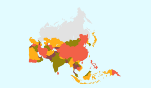
3. Unveiling immune cell diversity across Asian populations
Kock et al., created the Asian Immune Diversity Atlas (AIDA), a five-nation single-cell reference of circulating immune cells from 619 healthy donors across Japan, South Korea, Thailand, India, and Singapore (including Chinese, Malay, and Indian populations). This expansive effort—analyzing 1,265,624 cells using 5′ scRNA-seq, B cell receptor sequencing (BCR-seq), T cell receptor sequencing (TCR-seq), and genotyping—revealed how self-reported population descriptors such as ethnicity, genetic ancestry, sex, and age influence the cellular and molecular properties of immune cells, which are critical for disease risk assessment, diagnostics, and personalized medicine.
The study uncovered significant demographic impacts on immune cell proportions. For instance, plasmacytoid dendritic cell (pDC) proportions decreased with age, B cells were more abundant in females, and natural killer (NK) cells were more abundant in males. Ethnicity-specific differences included elevated B cell proportions in Singaporean Malay donors, lower myeloid cell proportions in Thai donors, lower NK cell proportions in Singaporean Indian donors, and lower T cell proportions in Korean donors.
The analyses also helped identify population-specific functional variants, including cell-type-specific expression quantitative trait loci (eQTLs), that contextualize disease-associated genetic loci, providing insights into disease risk and pathogenesis. Enrichment analysis of transcriptomic neighborhoods revealed fine-grained trends, such as an enrichment of naive B cells in females, a depletion of CD8⁺ naive T cells in older individuals (≥50 years), and an enrichment of gamma-delta T cells in Singaporean Malay donors.
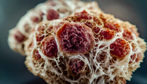
4. Decoding the tumor ecosystem of papillary thyroid carcinoma
This captivating study by Pu et al., published in Nature Communications, employed single-cell RNA sequencing (scRNA-seq) to profile 158,577 cells from 11 PTC patients, encompassing paratumors, localized/advanced tumors, lymph nodes, and radioactive iodine (RAI)-refractory metastases. This extensive analysis illuminated the cellular and molecular dynamics of PTC’s tumor ecosystem, providing insights into prognosis and therapeutic strategies.
The study pinpointed a ‘cancer-primed’ premalignant thyrocyte population that exhibited normal morphology but displayed altered transcriptomes, suggesting early malignant potential, though this finding, primarily based on one patient, requires further validation. Malignant thyrocytes were categorized into three phenotypes—follicular-like, partial-epithelial-mesenchymal-transition-like, and dedifferentiation-like—whose proportions shaped tumor subtypes, characteristics, and RAI responses.

A key discovery was the BRAF-like-B subtype, characterized by dedifferentiation-like thyrocytes and abundant cancer-associated fibroblasts, which correlated with poorer prognosis yet showed potential for immunotherapy. Additionally, the researchers identified vascular-immune crosstalk, particularly involving tip endothelial cells, supporting the potential of combined anti-angiogenic and immunotherapy approaches.
5. Decoding the complex ecosystem of diffuse large B cell lymphoma
This study by Steen et al., published in Cancer Cell, mapped the tumor microenvironment (TME) and malignant B cell states in diffuse large B cell lymphoma (DLBCL), the most common hematologic malignancy. Using their novel EcoTyper framework, which integrates bulk transcriptomic deconvolution via CIBERSORTx with single-cell RNA sequencing (scRNA-seq), the team analyzed 1,577 DLBCL tumors across four cohorts and profiled 173,028 single cells from lymphoid tissues, including four de novo DLBCL tumors, three follicular lymphomas (one undergoing transformation), one pediatric tonsil, and five publicly available scRNA-seq datasets, using 5’ scRNA-seq and scVDJ-seq.
The study identified 44 distinct cell states across 13 cell types, including five malignant B cell states with unique prognostic and differentiation profiles, validated in scRNA-seq datasets and mapped to normal B cell phenotypes such as germinal center B cells, pre-memory B cells, and pre-plasmablasts. For instance,
- State S1, enriched in germinal center B cell-like (GCB) DLBCL, predicted favorable overall survival (OS)
- State S2, with mixed COO representation, also independently predicted OS, highlighting additional prognostic heterogeneity
- State S5, associated with activated B cell-like (ABC) DLBCL, correlated with poorer OS across four cohorts
In addition, the study also revealed nine lymphoma ecotypes (LEs)—multicellular communities defined by co-occurrence patterns of cell states—that extended beyond conventional COO and genotypic classifications. Spatial transcriptomics using 10x Visium, applied to a normal human lymph node, further validated the spatial co-localization of cell states within LEs, confirming patterns such as the enrichment of B cell state S1 in B cell follicles and the proximity of CD8 T cell states to follicular structures.
6. Charting cell states and tumor microenvironments across human solid tumors
Luca et al., constructed a transcriptional atlas of cell states and ecosystems across 16 types of human carcinoma. Using their innovative EcoTyper framework, the team integrated bulk, single-cell, and spatial transcriptomics to analyze 5,946 tumor and 529 normal transcriptomes from The Cancer Genome Atlas (TCGA), alongside ~200,000 single-cell transcriptomes from breast, colorectal, head, and neck, and non-small cell lung cancers.
The study identified 69 transcriptionally distinct cell states across 12 major cell types, including immune, stromal, and epithelial cells. Most states were ubiquitous across tumor types, enriched in malignant tissue, and prognostic, with 39 states significantly associated with overall survival. Notably, M1 macrophages predicted favorable outcomes, while M2-like subsets, including a novel AEBP1+ foamy macrophage state, correlated with adverse survival.
The team further examined cell-state co-occurrence and defined 10 carcinoma ecotypes (CEs), which are multicellular communities with distinct biology and clinical implications. Of these, three ecotypes with myeloid and stromal dominance linked to poor prognosis, while two proinflammatory ecotypes were associated with early tumorigenesis. Techniques such as spatial transcriptomics and immunofluorescence validated the organization of these ecotypes, revealing ligand–receptor interactions that drive intercellular communication. Check out the full-text of the study here: https://www.sciencedirect.com/science/article/pii/S0092867421010618
Single-cell sequencing has emerged as a powerful tool in medical research, allowing scientists to investigate the inner workings of individual cells with exceptional clarity. Recognized as “Method of the Year” by Nature in 2013, the technology goes beyond average signals to uncover the intricate complexity of cellular behavior. They capture the transcriptomes of single cells, exposing the rare cell types and states that would otherwise remain undetected by bulk approaches. The resulting high-resolution cellular maps help dissect cell populations and uncover complex interactions, offering new insights into the regular physiological processes and disease mechanisms. With a bunch of applications spanning across therapy areas such as oncology, immunology, reproductive health, infectious diseases, and neurology, this technology is poised to drive the next wave of diagnostic and therapeutic breakthroughs.
About DataPrudence’s Single-Cell Sequencing Data Analysis
At DataPrudence, we are here to help you get the most out of your single-cell omics data. We offer end-to-end support, from high-quality preprocessing and integration across data types to delivering results that meet the standards of top-tier publications.
Whether you are mapping complex cancer cell populations or decoding disease pathways, we provide the expertise and tools needed to move your research forward. Reach out to us today and discover how we can help turn your data into discovery.